INTRODUCTION:
Odontogenic sinusitis is a distinct and severe form of inflammation of the maxillary sinus, which originates from dental conditions affecting the sinus.
Due to the anatomical proximity of the upper jaw to the floor of the maxillary sinus, it is usually caused by the posterior teeth of the upper jaw (from the second premolar backwards), whose roots communicate with the maxillary sinus.
If these teeth are non-vital, or if there are cysts originating from root-treated or damaged teeth, they may transmit bacteria from the teeth (very often anaerobic bacteria) into the maxillary sinus.
This is odontogenic sinusitis, which can often be detected by dentists and treated effectively.
The treatment of odontogenic sinusitis requires a combined approach, the most important part of which is endoscopic surgery - F.E.S.S. The affected tooth or cyst must be removed, and at the same time, any communication that may exist with the maxillary sinus must be closed.
CASE PRESENTATION:
A middle-aged female patient with a history of rejection of dental implants in the left upper jaw was referred by her Maxillofacial Surgeon with a CT scan of the paranasal sinuses showing complete opacification of the left maxillary sinus (antrum) and the left anterior ethmoid sinus.
The patient reported no history of rhinitis or sinusitis and presented with no other symptoms.
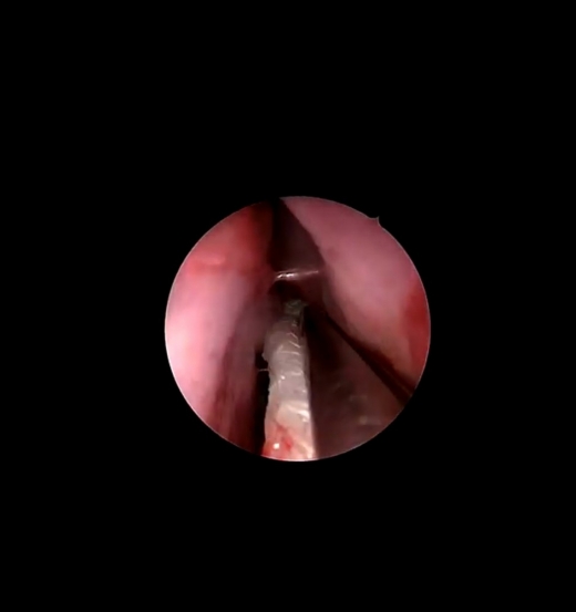
During endoscopic examination of the left middle nasal meatus at the clinic, a solid mass of polypoid texture was found protruding through the opening of the left maxillary sinus.
It was immediately decided that the patient would undergo endoscopic surgery to remove the polypoid masses from the paranasal sinuses (F.E.S.S.) at the Athens ENT Clinic.
Specifically, under endoscopic guidance, a permanent and wide middle meatal antrostomy was performed, that is, widening of the natural opening of the maxillary sinus. The polyp mass was removed, while the pathology within the affected maxillary sinus was simultaneously cleared. An anterior ethmoidectomy was also performed to remove the polypoid formations from the left anterior ethmoid air cells.
A total of 10 polypoid tissue fragments, each measuring between 0.3 and 1 cm in diameter, were removed and sent for histopathological examination - biopsy.
BIOPSY:
Microscopic examination of all tissue fragments showed that they consisted of portions of inflammatory nasal polyps, mostly covered by respiratory-type epithelium with mild hyperplastic changes and containing edematous stroma with areas of cystic degeneration.
Areas of granulation tissue were also observed, characterized by chronically inflamed stroma containing lymphocytes, histiocytes and plasma cells, areas of fibrosis and focal sclerosis of the interstitial tissue, as well as areas of hyperplastic mucus-producing glandular formations.
The findings are consistent with reactive tissue that may develop following irritation of the mucosa of the maxillary sinus by a foreign body (e.g. an implant).
Are you looking for answers and seeking a solution to your health concerns?
Monday to Thursday: 5 pm - 8 pm and Friday: 10:30 am - 1 pm
Call usOR
Please fill out the contact form: