A 32-year-old male patient, an amateur spearfisherman, non-smoker and with no history of allergic rhinitis, presented to the clinic with a CT scan showing a localized polypoid formation, measuring 1 x 1.5 cm, inside the right frontal sinus, in contact with its posterior wall.
Although the patient had no previous history of sinusitis, he had been experiencing progressively worsening frontal headaches over the previous two months, and was no longer able to dive as he used to. Endoscopic examination at the clinic revealed no significant findings apart from a deviated nasal septum and hypertrophic nasal turbinates.
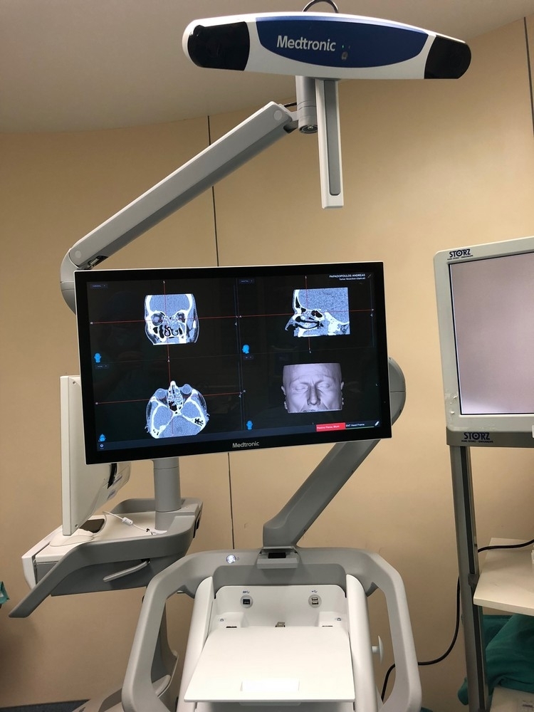
Due to the very difficult anatomical location of the polyp and the close anatomical relationship between the frontal sinus and the brain, it was decided to perform endoscopic removal using an image-guided navigation system - Navigator.
The patient was also asked to undergo a special type of preoperative CT scan of the facial skeleton, adapted for the navigation system, in order to prepare and plan the surgery in the best possible way.
The procedure took place at the Athens ENT Clinic and was completed with absolute success in approximately 2 hours.
In addition to the endoscopic removal of the polyp from the frontal sinus using the image-guided navigation system (Navigator), the natural opening of the frontal sinus was also widened in order to prevent future recurrence of the pathology.
At the same time, surgery was performed to correct the deviated nasal septum and reduce the hypertrophic nasal turbinates. The patient experienced not the slightest pain after surgery and was discharged the very next morning.
Thanks to the modern endoscopic approach to the frontal sinus, there were no external incisions, scars or marks on the face, nor any bruising or swelling above the nose, on the forehead or around the eyebrow. His postoperative course was completely uneventful.
After surgery, the patient underwent skin prick tests (Prick Test) to detect a possible allergic trigger, but the results were normal.
Up to 1.5 years after surgery, the patient remains free of disease recurrence. He is monitored regularly at the clinic, and endoscopic examinations show that the opening of his frontal sinus remains patent. Finally, he can comfortably dive to great depths without pain or any other discomfort.
Extensive excerpts from the procedure are shown below:
Although the patient had no previous history of sinusitis, he had been experiencing progressively worsening frontal headaches over the previous two months, and was no longer able to dive as he used to. Endoscopic examination at the clinic revealed no significant findings apart from a deviated nasal septum and hypertrophic nasal turbinates.
Due to the very difficult anatomical location of the polyp and the close anatomical relationship between the frontal sinus and the brain, it was decided to perform endoscopic removal using an image-guided navigation system - Navigator.
The patient was also asked to undergo a special type of preoperative CT scan of the facial skeleton, adapted for the navigation system, in order to prepare and plan the surgery in the best possible way.
The procedure took place at the Athens ENT Clinic and was completed with absolute success in approximately 2 hours.
In addition to the endoscopic removal of the polyp from the frontal sinus using the image-guided navigation system (Navigator), the natural opening of the frontal sinus was also widened in order to prevent future recurrence of the pathology.
At the same time, surgery was performed to correct the deviated nasal septum and reduce the hypertrophic nasal turbinates. The patient experienced not the slightest pain after surgery and was discharged the very next morning.
Thanks to the modern endoscopic approach to the frontal sinus, there were no external incisions, scars or marks on the face, nor any bruising or swelling above the nose, on the forehead or around the eyebrow. His postoperative course was completely uneventful.
After surgery, the patient underwent skin prick tests (Prick Test) to detect a possible allergic trigger, but the results were normal.
Up to 1.5 years after surgery, the patient remains free of disease recurrence. He is monitored regularly at the clinic, and endoscopic examinations show that the opening of his frontal sinus remains patent. Finally, he can comfortably dive to great depths without pain or any other discomfort.
Extensive excerpts from the procedure are shown below:
Are you looking for answers and seeking a solution to your health concerns?
Monday to Thursday: 5 pm - 8 pm and Friday: 10:30 am - 1 pm
Call usOR
Please fill out the contact form: