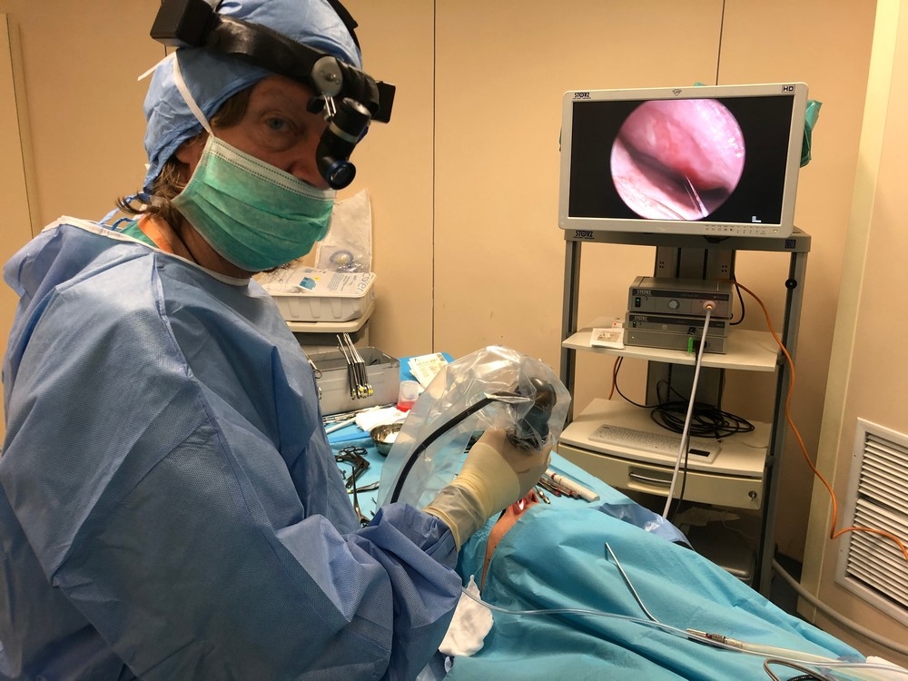
Treatment of a Patient with a Fungal Ball of the Sphenoid Sinus - Endoscopic Endonasal Transsphenoidal Removal
A 48-year-old female patient presented to Dr. Fotopoulos's clinic with persistent migraine-like headaches for at least 1 year.
The patient also complained of almost constant nasal breathing difficulty, with impaired airflow through the nose.
The patient had received multiple courses of antibiotics in the recent past, without any clinical improvement. She had no history of diabetes mellitus or immunosuppression. During the clinical examination at the clinic, nasal endoscopy revealed no significant findings apart from marked deviation of the nasal septum and hypertrophy of the nasal turbinates.
The patient was referred for a CT scan of the nose and paranasal sinuses, including coronal sections.
The CT scan showed complete opacification of the right sphenoid sinus by dense material, consistent with the presence of a fungal ball. At the same time, deviation of the nasal septum and hypertrophy of the inferior nasal turbinates were also confirmed.
All the other paranasal sinuses were free of pathology.
It was decided that the patient would undergo surgery at the Athens ENT Clinic for removal of the fungal ball from the sphenoid sinus using endoscopic microsurgery, with the removed material being sent for biopsy.
Indeed, the endoscopic procedure - F.E.S.S. - was performed on the right sphenoid sinus, and the fungal mass was completely removed under microscopic camera guidance. The material was sent to the pathology laboratory for histopathological examination.
At the same time, in addition to carefully clearing the sphenoid sinus of the fungal mass, its opening was permanently widened to improve communication with the nasal cavity and, consequently, improve ventilation of the sphenoid sinus.
In addition, the patient's deviated nasal septum was corrected and the hypertrophic nasal turbinates were reduced in order to restore nasal breathing.
The patient was discharged on the same day without any particular problems. Seven days later, the biopsy report showed: "Markedly compacted clots consisting of colonies of fungal hyphae (fungal ball) within mucous or hyphae-free material (PAS-positive staining)."
The patient is recovering uneventfully and did not require any local or systemic antifungal treatment. A full immunological assessment and frequent follow-up examinations with nasal endoscopy and MRI scans were recommended.
What is a fungal ball of the paranasal sinuses? What symptoms does it cause and how is it treated?
A fungal ball of the paranasal sinuses is a benign inflammatory condition that may mimic a tumor on imaging studies and can be successfully treated with endoscopic surgery.
Suitable conditions for their development include moisture, warmth and darkness, all of which are present in the paranasal sinuses. Excessive use of antibiotics, which disrupts the normal flora, as well as the increasing use of immunosuppressive medications, are factors that favor fungal growth.
Studies have shown that dental surgical procedures in the oral cavity also play an important role in the pathogenesis of fungal balls.
More specifically, it has been observed that a fungal ball may develop following tooth extraction, periodontal destruction or, more commonly, endodontic treatment.
Clinically, a fungal ball appears as a solid mass and may be misleading, often giving the impression of a tumor.
In cases of a fungal ball of the sphenoid sinus, headache (mainly retro-orbital, but also frontal or temporoparietal), postnasal drip and cough are the most common symptoms.
Much more rarely, the clinical presentation may include epistaxis (minor nasal bleeding), neurological symptoms such as visual disturbances, blurred vision, transient loss of vision, or even dysphonia.
The examination of choice is a CT scan of the facial skeleton. MRI is not necessary.
However, it is indicated in cases of sphenoid sinusitis and pansinusitis when there is bone erosion and possible contact with the contents of the eyeball or the structures of the brain must be ruled out.
Macroscopically, a fungal ball has a cheese-like consistency. It may be green, yellow, brown or black and can be easily detached from the mucosa. The postoperative course is monitored clinically and endoscopically through nasal endoscopy.
The treatment of choice is removal of the fungal ball through Functional Endoscopic Sinus Surgery (FESS), which allows widening of the opening of the affected sinus and removal of all fungal elements, which are sent to the pathology laboratory for biopsy.
No local or systemic antifungal treatment is required.
Are you looking for answers and seeking a solution to your health concerns?
Monday to Thursday: 5 pm - 8 pm and Friday: 10:30 am - 1 pm
Call usOR
Please fill out the contact form: